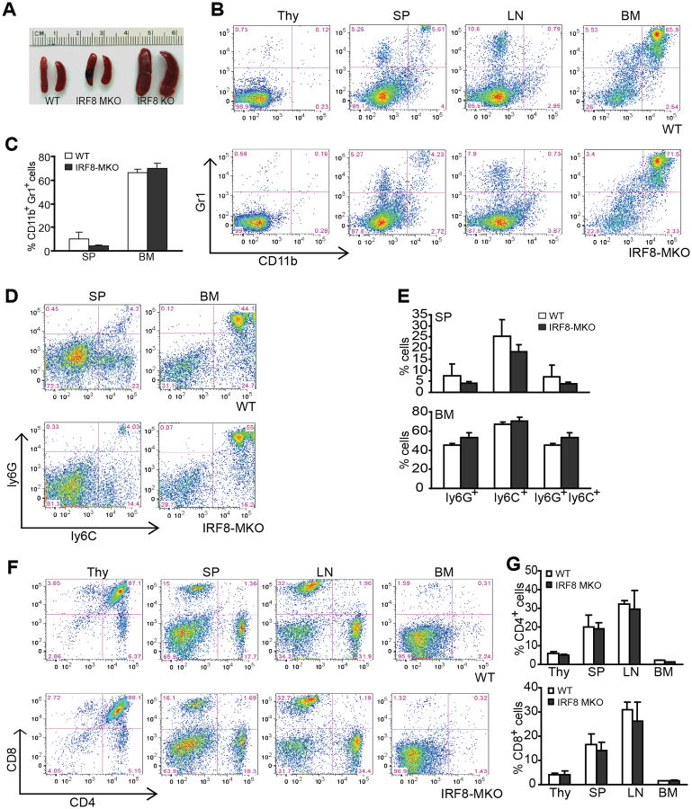
Figure 2. Mice with IRF8 deficiency only in myeloid cells exhibit normal myelopoiesis.
A. Spleen morphology of three month old WT, IRF8 MKO and IRF8 KO mice. B. Percentages of CD11b+Gr1+ MDSCs in thymus (Thy), spleen (SP), lymph node (LN) and bone marrow (BM) of WT and IRF8 MKO mice. Shown are representative results from one mouse of three mice. C. Quantitation of CD11b+Gr1+ in SP and BM of WT (n=3) and IRF8 MKO (n=3) mice as shown in B. D. Subsets of Gr1+ (Ly6C+ and Ly6G+) myeloid cells SP and BM. Shown are representative results from one mouse of three mice. Column: mean, bar: SD. E. Quantification of subsets of Gr1+ myeloid cells in SP and BM of WT (n=3) and IRF8MKO (n=3) mice. F. The indicated tissues were collected from WT and IRF8 MKO mice, stained with the CD4- and CD8-specific mAbs. Shown are representative images CD4+ and CD8+ T cell profiles. G. Quantification of CD4+ and CD8+ T cells in the indicated tissues from WT (n=3) and IRF8 MKO (n=3) mice. Column: mean, bar: SD.

