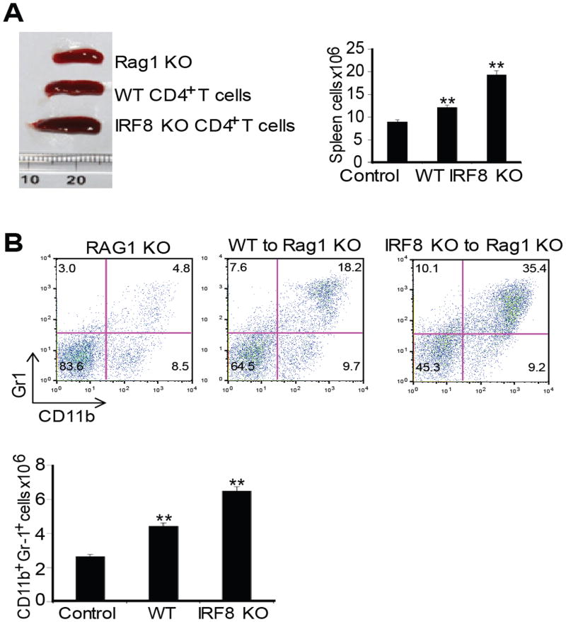
Figure 5. IRF8-deficient T cells induce MDSC accumulation.
A. CD4+ T cells were purified from WT and IRF8 TKO mice and adoptively transferred to Rag1 KO mice. Mice were analyzed 14 days after cell transfer. Spleen morphology (left panel), and total spleen cells (right panel) are shown. B. MDSC profiles in the Rag1 KO mice, Rag1 KO mice that have received WT T cells, and Rag1 KO mice that have received IRF8 KO T cells as shown in A. Spleen cells were stained with H2Kb-, CD11b- and Gr1-specific mAbs. H-2Kb+ cells were gated and analyzed for CD11b+Gr1+ cells. Shown are representative results of one of three mice (top panel). MDSCs in each type of mice were quantified and presented at the bottom panel. Column: mean, bar: SD. ** p<0.01.

