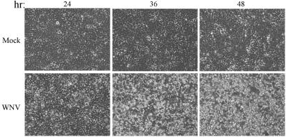
FIG. 2.
WNV-induced CPE. 293 cells were either mock treated or infected with WNV at an MOI of 5 and then incubated at 37°C for the time in hours (hr) indicated above each panel. Virus-induced CPE presents as a distortion of the cell monolayer and was visualized by using a Zeiss light microscope. The images were captured with a digital camera.

