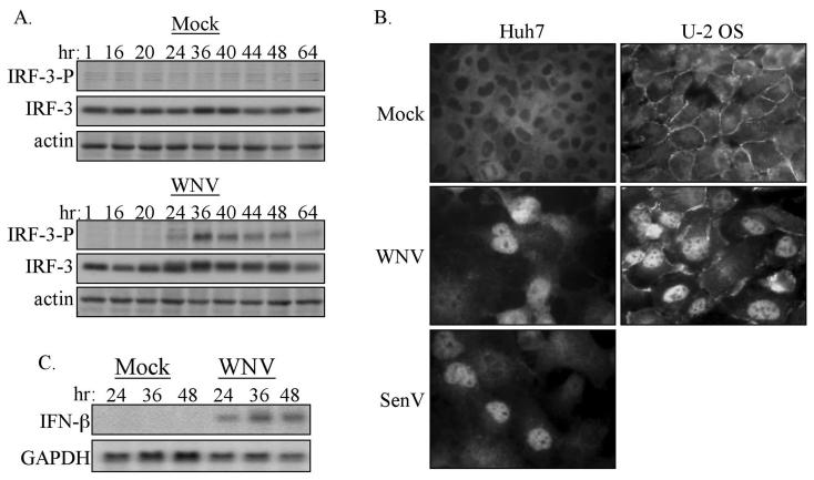
FIG. 6.
Characterization of the activation state of IRF-3 in WNV-NY-infected cells. (A) Phosphorylation state of IRF-3. Whole-cell lysates were recovered from mock- and WNV-NY-infected 293 cells over a 64-h time course, and immunoblot analysis was performed with an antibody specific for the phosphoserine 396 isoform of IRF-3 (IRF-3-P) (upper panels). Blots were stripped and reprobed with anti-sera against total IRF-3 (middle panels) or actin (lower panels). (B) IRF-3 localization in mock-, WNV-, or SenV-infected Huh7 or U-2OS cells was detected by indirect immunofluorescence analysis with IRF-3 polyclonal antiserum and a fluorescein isothiocyanate-conjugated secondary antibody. Images were acquired by using a Ziess Axiovert fluorescence microscope equipped with a digital camera and Axiovision software. (C) Induction of IFN-β expression by WNV-NY. IFN-β and GAPDH expression in mock- or WNV-NY-infected 293 cells was assessed by Northern blot analysis of total RNA harvested at the indicated times postinfection.

