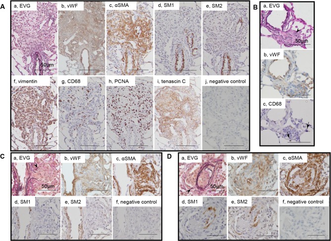
Fig 3. Myofibroblasts in plexiform lesions.
Photomicrographs of serial sections of complex plexiform lesions in a rat 13 weeks after initial treatment (Panels A) and sprouting plexiform lesions (Panels B, C, D). Immunohistochemical findings using antibodies for various antibodies were presented. αSMA+, vimentin+ supporting cells, which underlies endothelial monolayers, were regarded as myofibroblasts and were negative for SM1 or SM2. PCNA indicates proliferating cell nuclear antigen. An arrow head indicates fragments of elastic laminae. Abbreviations are described in Fig. 2.

