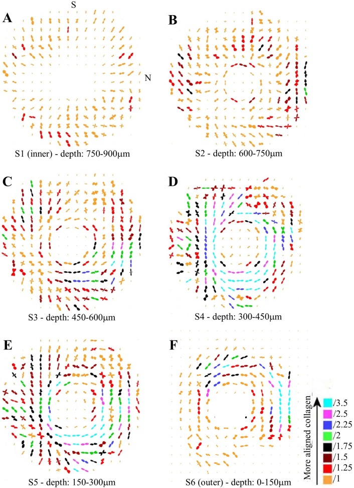
Fig 4. Collagen orientation maps.
Representative polar vector maps of preferential collagen fibril orientation across six serial sections (S1-S6) of human peripapillary sclera, at the stromal depths indicated. The inner section refers to that bordering the choroid, whereas the outer refers to that adjacent to the episclera. The plots have been scaled according to the color key and the superior (S) and nasal (N) aspects are indicated. Sampling interval: 0.5mm.

