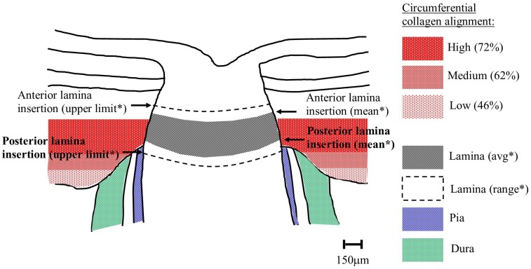
Fig 7. Comparison of circumferential collagen and lamina insertion depths.
Schematic of human optic nerve and peripapillary sclera cross-section, showing the location of the circumferential scleral collagen, as determined by WAXS, in relation to the typical position of the lamina cribrosa in a generic middle-aged/elderly normal human eye. % circumferential alignment is expressed as mean anisotropy, averaged over the four quadrants of eight eyes. *Distances of lamina insertion sites (average and range) are taken from the literature[20–22, 26].

