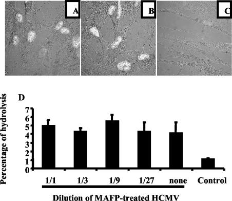
FIG. 9.
Inhibition of infection by MAFP is due to specific inhibition of HCMV-borne, but not cell-borne, cPLA2. MRC5 cells were seeded on glass coverslips in six-well plates (2.5 × 10⁵/well), deprived of FCS for 16 h, and incubated with a mixture of HCMV plus MAFP-treated HCMV (A), untreated HCMV alone (B), and MAFP-treated HCMV alone (C) (AD169, MOI = 0.1) for 1 h at 37°C. Unadsorbed virus was removed with PBS, and cells were further cultured for 5 days. Expression of IE1 and IE2 was analyzed by confocal microscopy (original magnification, ×60). (D) MRC5 cells were seeded in 24-well plates, deprived of FCS for 16 h, and then incubated for 2 h with dilutions of HCMV (original MOI = 0.1) treated with MAFP or not treated. The control represents cells incubated with MAFP only. Cells were washed, harvested, and lysed by sonication. Lysates were incubated for 1 h with BODIPY-PC, and lipid extraction and HPLC analysis of fluorescent metabolites were performed as described in Materials and Methods.

