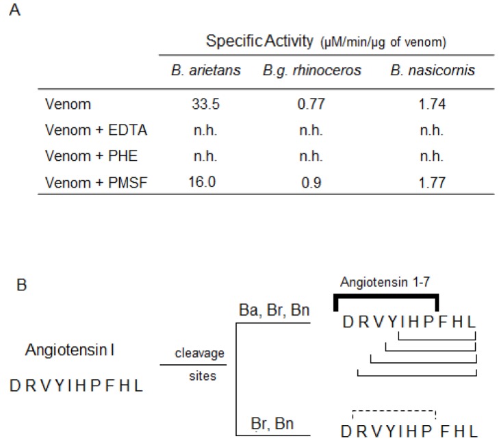
Fig 5. Cleavage of Angiotensin I by Bitis ssp venoms and identification of the cleavage sites on the primary sequence.
[A] Angiotensin I (65 μM) was incubated at 37°C with 1 μg of Bitis arietans venom (1 h/37°C) or 5 μg of B. nasicornis or B. g. rhinoceros venoms (2 h/37°C) in phosphate buffer (50 mM sodium phosphate, 20 mM NaCl, pH 7.4). In parallel, the venoms were pre-incubated, 30 min prior the addition of angiotensin I, with PHE (5 mM), PMSF (5 mM) or EDTA (100 mM). [B] The hydrolysis products, collected during the reverse-phase chromatography, were submitted to mass spectrometry analysis. All venoms cleaved angiotensin I in different cleavage sites. The lines indicate the cleavage sites on angiotensin I primary sequence after treatment with the different Bitis venoms. The bold solid line indicates the cleavage point for angiotensin 1–7 generation and the solid lines indicate the other cleavage points observed after the treatment with the venoms. The dashed line indicates the sites on angiotensin I primary sequence cleaved only by B. g. rhinoceros and B. nasicornis venoms.

