Fig. 1.

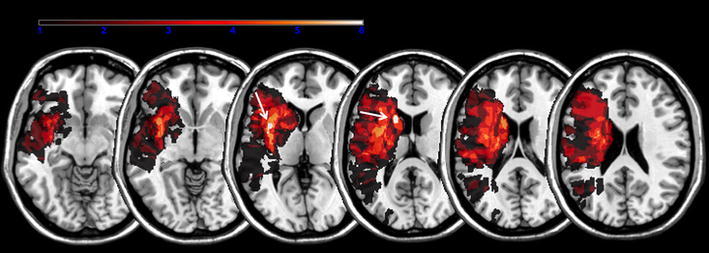
Lesion maps. Lesion overlay maps incorporating seven patients with common lesions in the insula (arrow MNI: x = −37, y = 7, z = 5) and seven in the caudate head (arrow MNI: x = −17, y = 14, z = 15) associated with abnormal yawning

Lesion maps. Lesion overlay maps incorporating seven patients with common lesions in the insula (arrow MNI: x = −37, y = 7, z = 5) and seven in the caudate head (arrow MNI: x = −17, y = 14, z = 15) associated with abnormal yawning