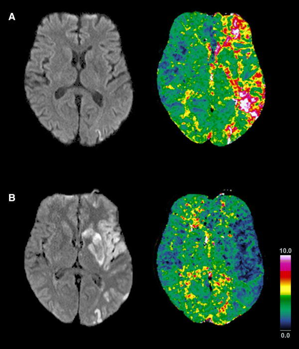
Fig. 2.

Abnormal yawning without initial DWI restrictions. (a) Evolution of the penumbra in a patient with abnormal yawning initially not related to DWI lesions in the caudate head or insula (Pat No 4). While cortical DWI restrictions were initially restricted to the frontal lobe (not shown) and parietal lobe, perfusion imaging revealed a widespread penumbra along the left MCA encompassing the insula and caudate head (TTP delay >4.5 s). (b) Follow-up after 48 h revealed prolonged infarction of the tissue at risk in the left insula, striatum and frontal and parietal lobe, now including the caudate head and the insula, with luxury perfusion of the infarcted tissue
