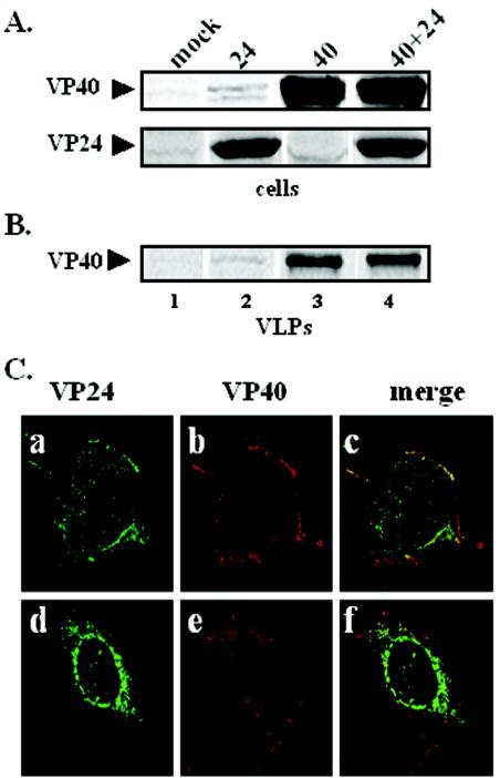
FIG. 2.
Functional budding assay and intracellular localization of VP40 and VP24. 293T cells were transfected with pCAGGS vector alone (lane 1), VP24 (lane 2), VP40 (lane 3), or VP24 plus VP40 (lane 4). VP40 and VP24 proteins were detected in cells (A), and VP40 was detected in budding VLPs (B) by immunoprecipitation and SDS-PAGE analysis at 30 hpt. (C) Cos-1 cells were cotransfected with VP40 and VP24, and fixed cells were visualized by confocal microscopy 24 hpt. VP24 is shown in green (panels a and c), VP40 is shown in red (panels b and e), and the merged images are shown in panels c and f. Yellow indicates colocalization.

