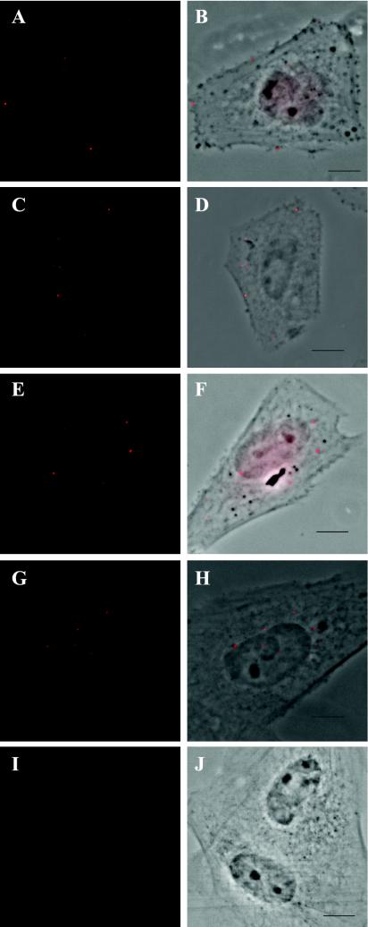
FIG. 1.
A time course of the progression of labeled HCMV into the nucleus. Human FFs were seeded onto glass coverslips, infected at an MOI of 1 by using protocol 1, harvested and stained for BrdU as indicated in the text (rat anti-BrdU Ab OBT030CX from Accurate Chemicals Scientific Corp., anti-rat tetramethyl rhodamine isothiocyanate-labeled secondary Ab from Jackson Immunoresearch Laborato-ries). Cells were analyzed on a Nikon Eclipse E800 epifluorescence microscope equipped with a digital camera and Metamorph imaging software. BrdU staining (red in Panels A, C, E, G, and I) was overlaid onto phase-contrast images in panels B, D, F, H, and J. (A and B) Cells incubated with virus on ice for 30 min prior to harvesting. (C and D) Cells harvested at 15 min post-cold release. (E and F) Cells harvested at 30 min post-cold release. (G and H) Cells harvested at 1 h post-cold release. (I and J) Cells incubated with supernatant gathered from mock-infected cells after treatment for 3 days with BrdU. Cells were harvested at 30 min p.i. Bar = 5 μm for all figures.

