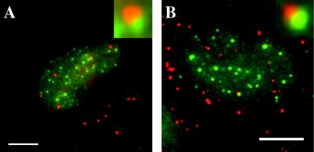
FIG. 3.
A subset of BrdU foci is juxtaposed to PML sites in infected nuclei. Cells were infected at an MOI of 3 using protocol 2, harvested at 3.5 h p.i., and stained for PML (green label) using Santa Cruz Ab SC966 detected with goat anti-mouse IgG1 Alexa Fluor-coupled secondary Ab (Molecular Probes). After a second round of fixation, cells were stained for BrdU as described in the text (red label). The small insets are further magnification of the overlapping regions within the nuclei pictured. (A) Cells were washed after 30 min p.i. (B) Cells were unwashed for the duration of the experiment.

