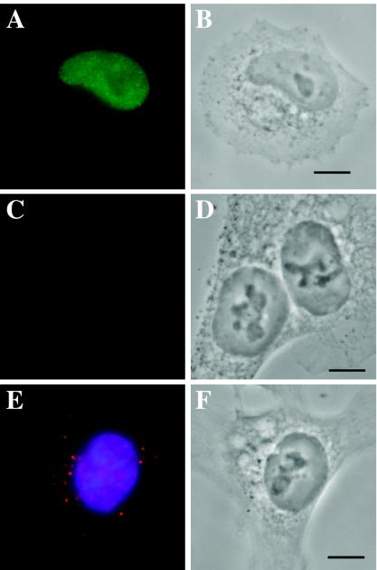
FIG. 4.
HCT116 cells are blocked for nuclear import. HCT116 cells were seeded onto polylysine-coated coverslips, infected at an MOI of 1 using protocol 2, and harvested at 24 h p.i. as described. All images were taken by using Z series. (A) FS2 cells stained for immediate-early protein IE1 using Ab p63-27 (a gift from William Britt, University of Alabama) followed by IgG2A anti-mouse Alexa Fluor-conjugated secondary Ab (Molecular Probes). (B) Phase-contrast image of cells in panel A. (C) HCT116 cells stained for IE1. (D) Phase-contrast image of Panel C. (E) Overlaid image of HCT116 nuclei stained for BrdU (red label) and DNA (blue label). (F) Phase-contrast image of panel E.

