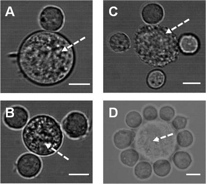
Figure 2.
Patterning of multiple cell types using holographic optical tweezers in co-cultures of: (A–B). Mouse embryonic and mesenchymal (arrow) stem cells. (C–D). Mouse primary calvarae cells (arrow) and embryonic stem cells. Imaging was achieved by use of integrated fluorescent and bright-field imaging system. Scale bar = 12 μm.

