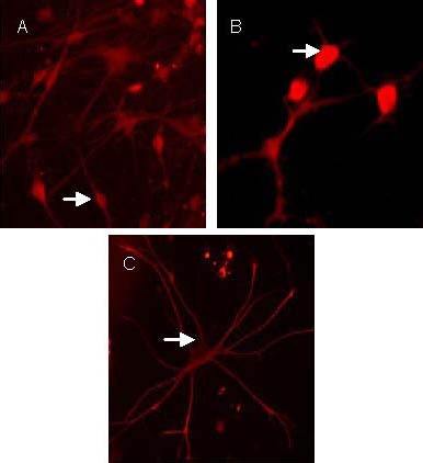
Figure 4.

Neonatal rat neural stem cells differentiated into neuron-specific enolase-positive neurons (A), 2’,3’-cyclic-nucleotide 3’-phosphodiesterase-positive oligodendrocytes (B), glial fibrillary acidic protein-positive astrocytes (C) after induction by 10% fetal bovine serum (Cy3, × 400).
Arrows show positive neurons. The positive neuron in Cy3 staining is red.
