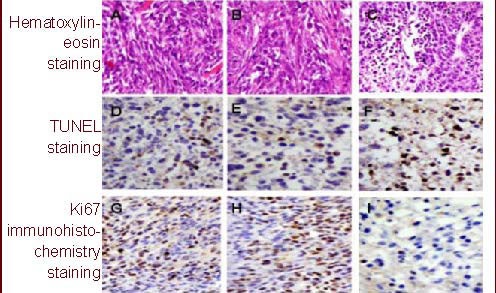
Figure 5.

Hematoxylin-eosin staining, terminal deoxynucleotidyl transferase-mediated dUTP-biotin nick end labeling and Ki67 immunohistochemistry analysis for rat glioma (× 100).
(A, D, G) In the untreated groups, the tumors grow very strong and apoptosis cells are little.
(B, E, H) In control vector group, apoptotic cells are also little, Ki67 positive cells are everywhere.
(C, F, I) In survivin small interfering RNA group, apoptotic cells increase significantly while Ki67 immuno-positive cells are reduced greatly. Positive cells: brown.
