Figure 1.

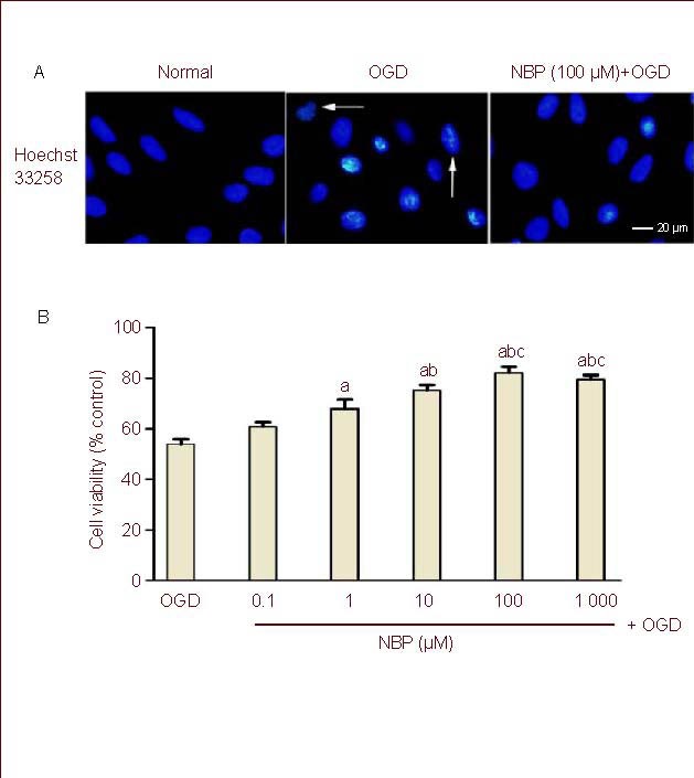
Effect of DL-3-n-butylphthalide (NBP) on cell damage induced by oxygen glucose deprivation (OGD) in rat brain microvascular endothelial cells (BMECs).
(A) BMECs were pretreatment with NBP (100 μM) for 1 hour, then underwent 2-hour OGD and 24-hour reperfusion.
BMEC nuclei were stained with Hoechst 33258. Crenation of nuclei and condensation of chromatin were evident in BMECs exposed to OGD (arrows) and OGD-induced nucleus morphological changes were obviously reduced in BMECs treated with NBP (100 μM) before exposure to OGD.
(B) Bar graph shows the effect of pretreatment with different concentrations of NBP on the viability of BMECs with OGD insult.
Each experimental group was expressed as percentage of that of the sham-OGD group (normal group).
Values were expressed as mean ± SEM. The results were obtained from five independent experiments performed in triplicate.
aP < 0.05, vs. the OGD group; bP < 0.05, vs. the NBP (1 μM) + OGD group; cP < 0.05, vs. the NBP (10 μM) + OGD group (one-way analysis of variance with the least significant difference post hoc test).
