
Figure 1.
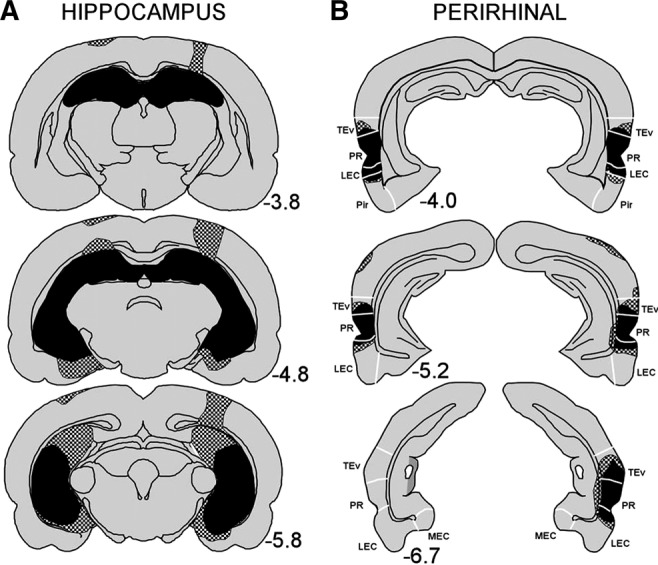
Reconstructions of coronal sections through the (A) hippocampus and (B) perirhinal cortex showing the smallest (black) and largest (stippled) lesion for the hippocampal and perirhinal lesion groups, respectively, in Experiment 1. Numbers represent the distance (millimeters) posterior to bregma. (TEv) ventral TE, (PR) perirhinal cortex, (LEC) lateral entorhinal cortex, (MEC) medial entorhinal cortex, (Pir) piriform area. White lines indicate approximate borders between these structures.