Figure 3.

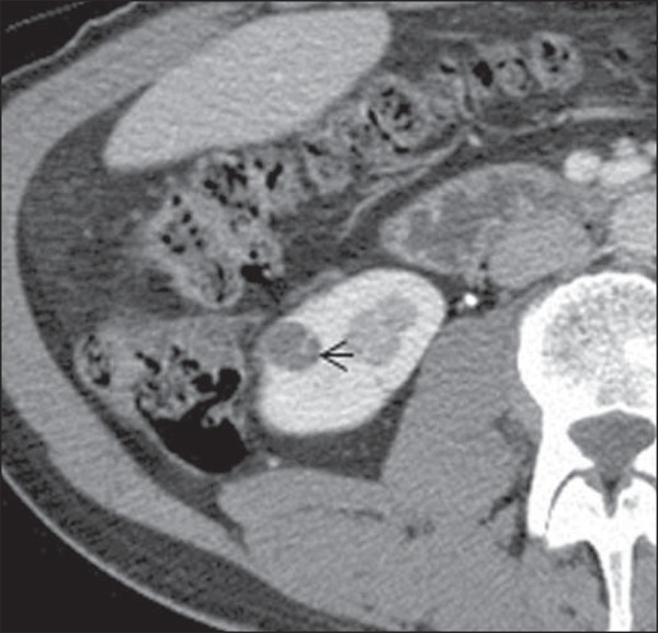
Bosniak II-F cyst. Contrast-enhanced CT image shows a partially exophytic cyst with a fine septation inside. Subtle nodularity is observed in the septum, which has perceptible but not measurable contrast-enhancement (arrow).

Bosniak II-F cyst. Contrast-enhanced CT image shows a partially exophytic cyst with a fine septation inside. Subtle nodularity is observed in the septum, which has perceptible but not measurable contrast-enhancement (arrow).