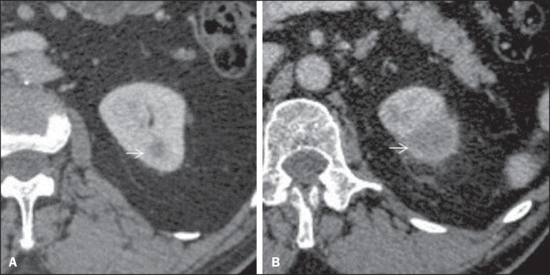
Figure 4.
Progression of a complex cystic lesion. A: Contrast-enhanced CT. Initial study shows a small hypodense cortical lesion in the upper pole of the left kidney (arrow). B: Contrast-enhanced CT image acquired four months later. Despite the significant enlargement of the lesion (arrow), it was classified as Bosniak II-F. After another follow-up scan that demonstrated further increase in dimensions, the lesion was resected and a clear cell renal cell carcinoma was diagnosed.

