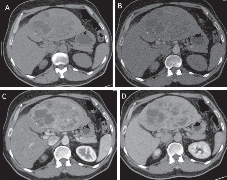
Figure 4.
Primary hepatic neuroendocrine carcinoma. Computed tomography, non-contrast enhanced image (A) and after intravenous contrast media injection (B,C,D) demonstrate the presence of a heterogeneous, quite vascularized mass with some foci of calcification and intermingled cystic areas, occupying the left liver lobe.

