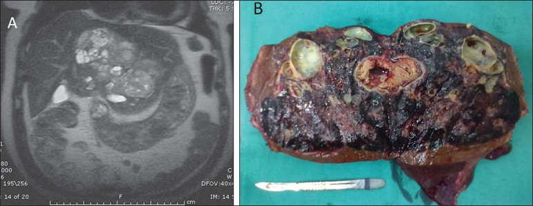
Figure 6.
Primary hepatic neuroendocrine carcinoma (same patient on Figures 4 and 5). MRI, coronal T2-weighted image (A) demonstrates heterogeneous mass occupying almost the whole left liver lobe, with hypersignal intermingled with multiple cystic areas, with excellent correspondence with the macroscopic aspect observed on the surgical specimen (B).

