Figure 1.

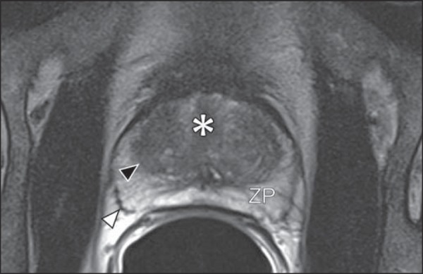
MR imaging of the normal prostate. T2-weighted sequence in the axial plane showing the prostate capsule (white arrowhead), the peripheral zone (ZP), the surgical capsule (black arrowhead) and internal gland (asterisk).

MR imaging of the normal prostate. T2-weighted sequence in the axial plane showing the prostate capsule (white arrowhead), the peripheral zone (ZP), the surgical capsule (black arrowhead) and internal gland (asterisk).