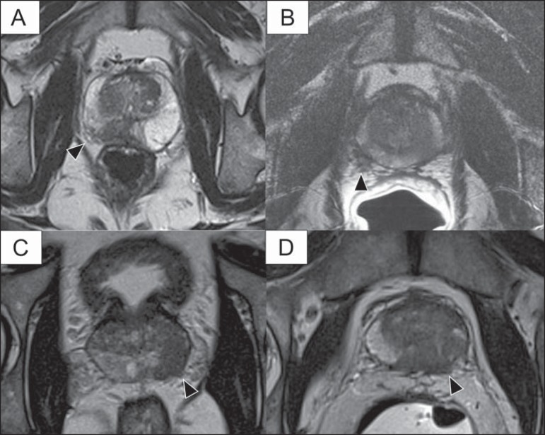
Figure 4.
T2-weighted MR images of the prostate, showing typical findings of extracapsular tumor extension marked by arrowheads in the following examples: asymmetry of the neurovascular bundle (A), tumor involvement of the neurovascular bundle (B), spiculated contour of the prostatic capsule (C), and focal bulging on the contour of the prostatic capsule (D).

