Figure 3.

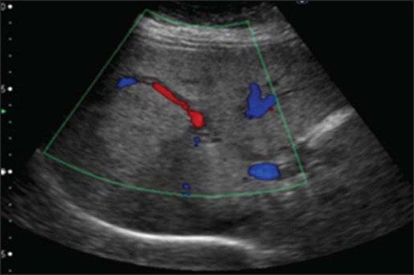
Angiomyolipoma. Ultrasonography demonstrates the presence of hyperechogenic liver mass with lobulated contours and central flow at Doppler, located in the segments VII/VIII.

Angiomyolipoma. Ultrasonography demonstrates the presence of hyperechogenic liver mass with lobulated contours and central flow at Doppler, located in the segments VII/VIII.