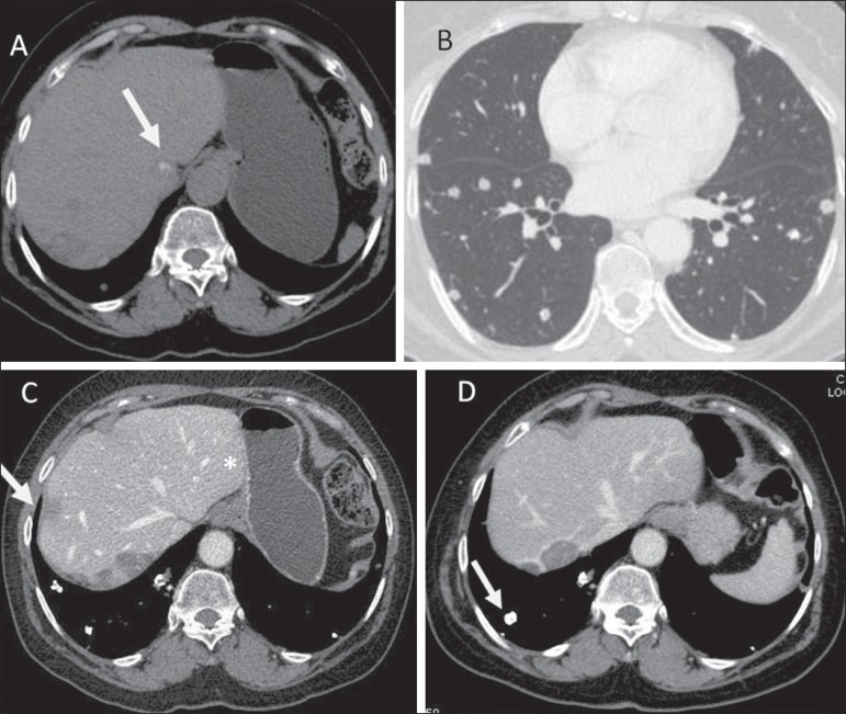
Figure 7.
Epithelioid hemangioendothelioma. Non-contrast-enhanced abdominal CT identifies the presence of hypodense peripheral hepatic nodules intermingled with foci of calcifications (arrow on A), which may occur in up to 25% of cases. Chest CT of the same patient (B) demonstrates the presence of sparse secondary nodules randomly distributed on the pulmonary parenchyma. At abdominal CT portal phase (C,D) hepatic nodules present with more intense enhancement in the periphery of the lesion, hepatic capsule retraction (arrow on C), besides calcified lung metastases (arrow on D).

