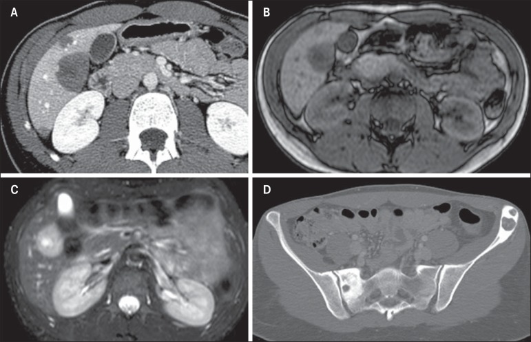
Figure 8.
Epithelioid hemangioendothelioma. A: Abdominal CT portal phase demonstrates the presence of hypovascular peripheral hepatic nodules. MRI demonstrates that the hepatic nodule presents low signal intensity at T1-weighted sequences (B) and high signal intensity at T2-weighted with target sign (C). At pelvic CT, insufflating osteolytic lesions are observed, with marginal sclerosis in the left iliac bone and scrum at right (D).

