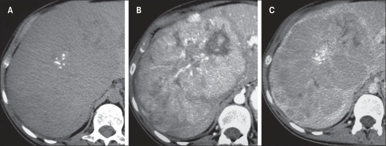
Figure 9.
A 23-year-old woman with no hepatopathy and negative tumor markers and with fibrolamellar hepatocellular carcinoma. Non-contrast-enhanced CT (A) arterial (B) and equilibrium (C) phases demonstrate the presence of a subtly hypodense large mass in the right liver lobe, with central punctiform calcifications presenting heterogeneous hypervascular enhancement with progressive appearance in its central region, intermingled with necrotic areas.

