Figure 5.
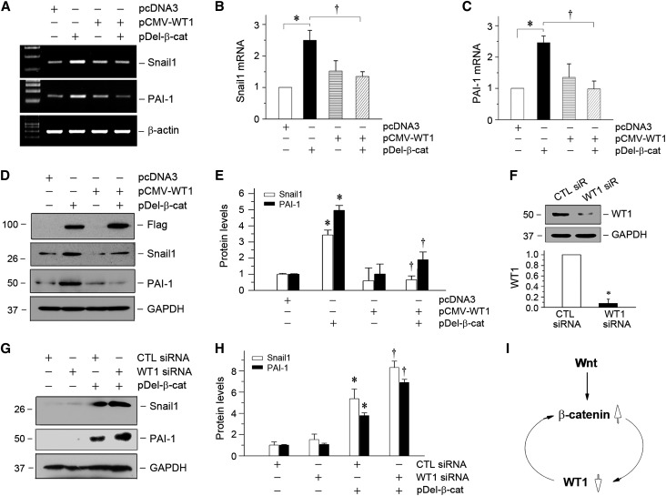
Loss of WT1 amplifies β-catenin signaling and augments its downstream gene expression in vitro. (A–C) RT-PCR analyses show that overexpression of WT1 abolished β-catenin–mediated gene expression. Mouse podocytes were transfected with WT1 expression vector (pCMV-WT1) or/and pDel-β-cat as indicated. The mRNA expressions of Snail1 and PAI-1 were assessed by RT-PCR. Representative (A) RT-PCR and quantitative data for (B) Snail1 or (C) PAI-1 are presented. *P<0.05 versus pcDNA3 controls; †P<0.05 versus pDel-β-cat alone (n=3). (D and E) Western blotting shows that overexpression of WT1 blocked the induction of β-catenin target genes in moue podocytes. (D) Representative Western blot was performed to analyze the expression of Snail1 and PAI-1. Expression of exogenous β-catenin was verified by assessing Flag. Quantitative data are presented in E. *P<0.05 versus pcDNA3 controls; †P<0.05 versus pDel-β-cat alone (n=3). (F) Knockdown of endogenous WT1 protein in podocytes. Mouse podocytes were transfected with control or WT1-specific siRNA, and WT1 protein expression was analyzed by Western blotting. Glyceraldehyde-3-phosphate dehydrogenase (GAPDH) was used to confirm equivalent loading. *P<0.05 versus siRNA (n=3). CTL, control; siR, siRNA. (G–I) Knockdown of endogenous WT1 augments β-catenin–mediated Snail1 and PAI-1 expression. Representative (G) Western blot and (H) quantitative data for Snail1 and PAI-1 are presented. *P<0.05 versus CTL siRNA; †P<0.05 versus pDel-β-cat plus CTL siRNA (n=3). (I) Diagram indicates that β-catenin activation and loss of WT1 constitute a vicious cycle, leading to podocyte dedifferentiation.

