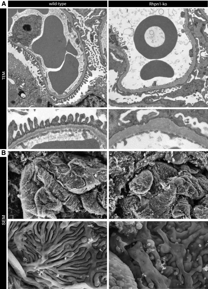
Figure 5.
Ultrastructural changes to podocytes and the GBM characterize the glomerular phenotype of Rhophilin-1 null mice. (A) Transmission electron microscopy (TEM) reveals widespread foot process effacement around capillary loops of Rhophilin-1 ko glomeruli. Also note that the basal surface of podocytes that juxtaposes the outer surface of the GBM shows membrane irregularities and invaginations. The GBM of ko mice is thickened, electron-dense, and homogeneous without clearly defined structural layers. (B) Scanning electron microscopy (SEM) images of the outer surface of capillary loops show a replacement of thin interdigitating podocyte foot processes of wild-type mice with flattened epithelial sheets and stubby foot processes. Microvillous transformation can also been seen on the apical surface of podocytes of Rhophilin-1 ko glomeruli. Magnification, ×10,000 in A, upper panel; ×25,000 in A, lower panel; ×7000 in B, upper panel; ×20,000 in B, lower panel.

