Figure 6.
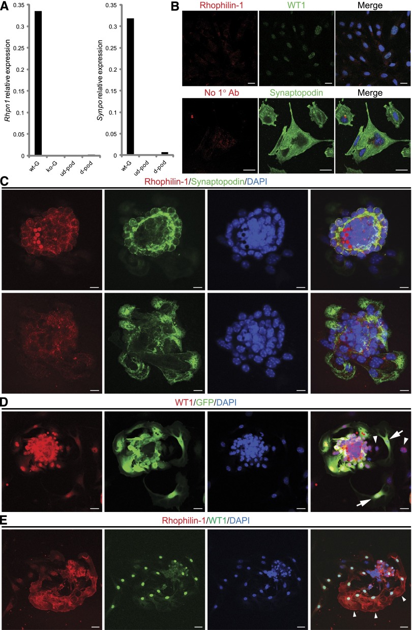
Rhophilin-1 expression in cell lines and primary cultures of glomerular explants. (A) Quantitative analysis of representative Rhpn1 gene expression in glomeruli isolated from wild-type (wt-G) and Rhpn1 null (ko-G) mice compared with undifferentiated (ud-pod) and differentiated (d-pod) cultured mouse podocytes. (B) Differentiation of cultured podocytes induces synaptopodin expression (lower panel) but does not elicit a similar increase in Rhophilin-1 expression (upper panel). Differentiated podocytes stain positively for WT1 and synaptopodin. Immunostaining in the presence or absence of Rhophilin-1 primary Ab gave similar background signal. Merge images are presented with 4′,6-diamidino-2-phenylindole (DAPI) nuclear counterstain. (C) Primary outgrown podocytes from wt glomeruli express synaptopodin but do not show significant Rhophilin-1 expression. Both Rhophilin-1 and synaptopodin are readily detected and colocalize in podocytes of the glomerular core (upper panel). Changing the plane of focus (lower panel) reveals outgrown primary podocytes that stain positive for synaptopodin but lack Rhophilin-1. (D) Primary cultures of glomeruli isolated from Rhpn1 null mice show podocyte dedifferentiation. Outgrown cells of the podocyte lineage that express WT1 can be found to costain with GFP (a reporter of Rhpn1 promoter activity; arrows). Some WT1-expressing outgrown podocytes have lost expression of GFP, indicating that the Rhpn1 promoter that drives GFP expression has been silenced and that significant dedifferentiation has occurred (arrowheads). (E) Endogenous Rhophilin-1 expression is observed in a subpopulation of primary outgrown podocytes that stain positively for WT1 (arrowheads). Scale bars, 20 μm in B, D, and E; 10 μm in C.

