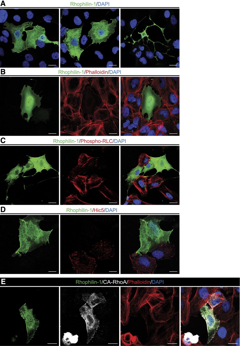
Figure 8.
Rhophilin-1 expression in human podocytes inhibits Rho-dependent signaling. (A) Heterologously expressed Rhophilin-1 partially localizes at the cell membrane and sites of cell–cell contact. (B) The actin stress fiber network (visualized with phalloidin) is disrupted in Rhophilin-1–expressing cells. (C) Rhophilin-1 inhibits phosphorylation of the RhoA substrate myosin RLC. (D) FAs (Hic5) are markedly reduced in Rhophilin-1–expressing cells. (E) Rhophilin-1 prevents CA-RhoA–dependent stress fiber formation and contractile phenotype. Note the smaller cell size and increased stress fiber formation in CA-RhoA–expressing cells compared with Rhophilin-1/CA-RhoA–expressing cells. DAPI, 4′,6-diamidino-2-phenylindole. Scale bars, 20 μm.

