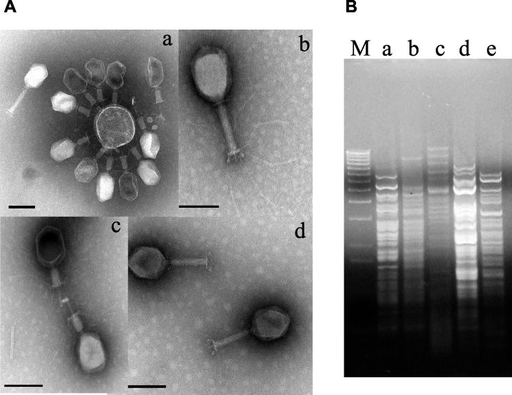
FIG. 1.
Four T4-like phages used in the mouse experiments. (A) Transmission electron microscopy picture of CsCl density gradient-purified bacteriophage JS4 (a), JSD.1 (b), JSL.6 (c), and JS94.1 (d). Negative staining was performed with uranyl acetate (c), ammonium molybdate (a and d), or phosphotungstic acid (c). The size bar corresponds to 100 nm. (B) Restriction analysis of phages (for lanes a to d, see corresponding subpanel in panel A; lane e, phage T4) with enzyme DraI. Lane M, DNA size marker (1-kb lambda DNA ladder; Invitrogen).

