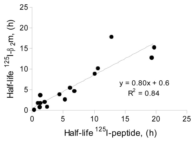
Figure 4.
Comparison of the dissociation rates measured with either 125I-β2m or 125I-peptide. Two different peptides (FLPSDYFPSV and RLPAYAPLL) were radiolabeled. pMHC’s were generated with different MHC-I molecules (A*02:01, A*02:02, A*02:03, A*02:04, A*02:05, A*02:11, A*02:19 and A*69:01) using either labeled peptide and excess of β2m or labeled β2m and excess peptide. For each pMHC the dissociation rate measured with labeled peptide was compared to the dissociation rate measured with labeled β2m.

