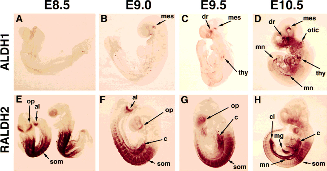
Fig. 4.
Localization of ALDH1 and RALDH2 proteins during stages E8.5–E10.5 of mouse embryogenesis. Embryos were subjected to whole-mount immunohistochemistry and prior to photography were cleared in benzyl alcohol:benzyl benzoate (1:2). Due to the transparent nature of the cleared embryos, both portions of bilateral structures are observed simultaneously. al, allantoic mesenchyme; c, coelomic cavity mesenchyme; cl, cloaca; dr, dorsal retina; mes, ventral flexure of mesencephalon; mg, midgut; mn, mesonephric ducts; op, optic vesicle/frontonasal mass; otic, otic vesicle; som, somite; thy, thymic primordia.

