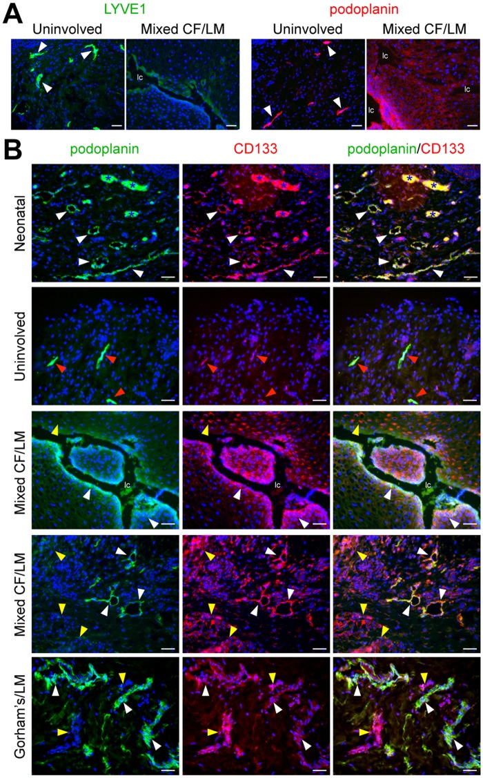
Fig 1. Identification of CD133+ cells in LMs of different subtypes and anatomical locations.
(A) LYVE1 and podoplanin staining of cervicofacial mixed LM tissue and patient-matched uninvolved tissue. White arrowheads mark normal lymphatics. (B) Podoplanin and CD133 staining of neonatal foreskin (postnatal day 1), uninvolved tissue, mixed cervicofacial (Mixed CF) LM tissues (2x), and Gorham’s dermal tissue. White arrowheads mark CD133+/podoplanin+ lymphatic endothelium. Red arrowheads mark CD133low/podoplanin+ lymphatic endothelium. Yellow arrowheads mark CD133+/podoplanin+ stromal cells. Blue asterisks mark blood vessels with autofluorescing red blood cells. Scale bars: 50μm. lymphatic channel (lc)

