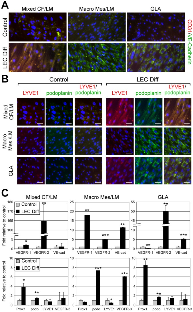
Fig 5. LMPCs differentiated into abnormal lymphatic endothelial cells.

LMPCs isolated from mixed cervicofacial (Mixed CF) LM, macrocystic mesenteric (Macro Mes) LM and a generalized lymphatic anomaly (GLA) were maintained in growth media (control) or lymphatic endothelial differentiation (LEC Diff) media for two weeks. (A) CD31/VE-cadherin and (B) LYVE1/podoplanin staining. Scale bars: 50μm. (C) VEGFR-1, VEGFR-2, VE-cadherin, Prox1, Podoplanin, LYVE1, and VEGFR-3 qRT-PCR of RNA isolated from LMPCs maintained in growth or LEC differentiation (LEC Diff) media. Data normalized to β-actin qRT-PCR and represented as mean ± s.e.m. * p < 0.05, ** p < 0.005, *** p < 0.0005.
