FIGURE 4:
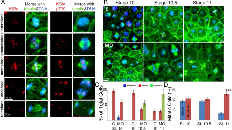
Kif2a localization and T70 phosphorylation during mitosis. Kif2a MO–treated caps at stages 10, 10.5, and 11 of development. (A) Confocal micrographs of interphase and mitotic cells in X. laevis animal caps double immunostained for Kif2a (red) and tubulin (green). DNA is stained blue with 4′,6-diamidino-2-phenylindole. For the phosphorylated form, the T70 protein is immunostained green and α-tubulin localized by a specific (DM1A) antibody red, and the DNA stains blue. During prophase and metaphase, Kif2a is localized to centromeres. During anaphase and telophase, Kif2a became localized to the spindle poles of the cell. The T70 phosphorylated form of Kif2a also localized to the centromeres during prophase and metaphase, and during anaphase, it is localized to the spindle. T-70 was also localized to the poles during metaphase, with slight localization at the poles during anaphase. In addition, pT70 exhibits strong localization to the midbody during telophase. (B) Single-cell embryos were microinjected with MilliQ H2O or the antisense MO for Kif2a (40 nl at 1 mg/ml) and allowed to develop in 0.3× MMR. At stages 10, 10.5, and 11, both groups were dissected, fixed, and stained for Kif2a (red), α-tubulin (green), and DNA (blue). There was an accumulation of multipolar spindles, with development within the Kif2a morphant groups between stages 10 and 11. (C) Quantification of cellular mitotic spindle types (bipolar vs. multipolar) within different, staged animal caps demonstrated an increase or accumulation of multipolar spindles after stage 10 within the MO-injected embryos compared with controls. No multipolar spindles were observed within controls or stage 10 MO-injected groups. (D) The percentage of animal cap cells that are undergoing mitosis and the percentage of mitotic cells within each phase of mitosis are illustrated for each stage and experimental group. ***p < 0.001. Error bars indicate SEM.

