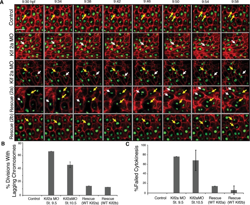
FIGURE 6:
Kif2a morphant phenotypes caused by lagging chromosomes and failure of cytokinesis. (A) Embryos were injected at two-cell stage with GAP43 RFP:RNA and H2B:GFP RNA (Control) and coinjected with Kif2a morpholino (Kif2aMO), MO plus human 2a RNA (Rescue 2a), or MO plus human 2b RNA (Rescue 2b). The animal caps were microdissected at stage 9 and placed in an imaging chamber for confocal microscopy. A time-lapse movie was made on a Zeiss 780 Confocal Microscope with the 25× objective and a framing rate of 30 s. Still frames of these movies are shown from the indicated time postfertilization. The morphant embryos display lagging chromosomes and failure of cytokinesis (indicated by white or yellow arrows). Scale bar, 20 μm. (B) From time-lapse movies taken from stages 9–12 (similar to those described earlier), quantification of divisions with lagging chromosomes was made for control embryos, Kif2a morphants at stage 9.5, Kif2a morphants at stage 10.5, and morphants rescued with either human Kif2a or Kif2b. Bars represent the percentage of divisions imaged with lagging chromosomes. n = 20 for each category of embryo. Control embryos displayed no divisions with lagging chromosomes. Error bars, SEM. (C) Percentage of divisions imaged with failed cytokinesis. n = 20 for the categories of embryos in B. Error bars, SEM.

