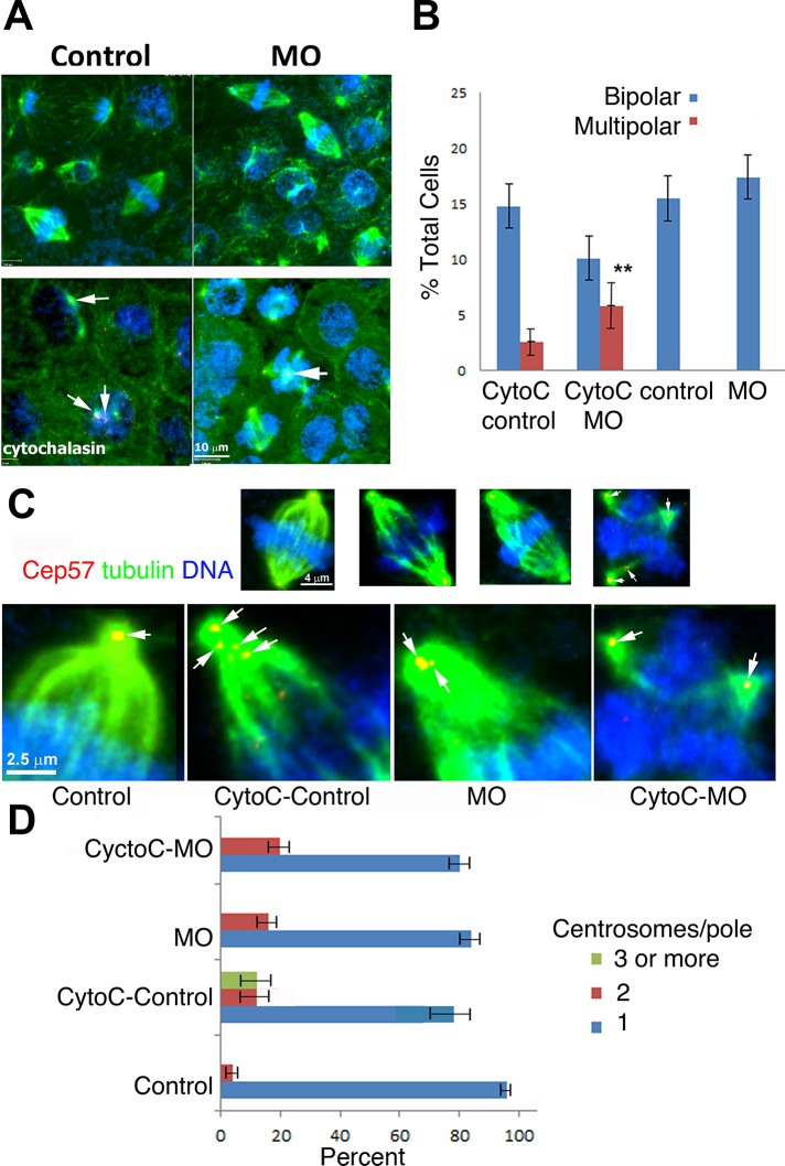
FIGURE 8:
Kif 2a-depleted cells poorly coalesce centrosomes. (A) Embryos were treated with 80 μM cytochalasin B at stages 8/9 and washed out of the drug after 1 h. At stage 10 embryos were fixed and imaged. Caps were immunostained for α-tubulin (green) and Cep57 (red) and stained for DNA (blue). Arrows (cytochalasin group) indicate centrosomes and multicentrosomes within cells. (B) Inhibition of cytokinesis generates multipolar spindles within stage 10 embryonic caps at a stage before their normal appearance in untreated MO-injected caps. Both cytochalasin-treated groups had multipolar spindles, but they were more abundant within the MO-injected group treated with DMSO instead of cytochalasin (control), depleted of Kif2a and DMSO (MO), control MO treated with cytochalasin B (CytoC control), or Kif2a-depleted embyros treated with cytochalasin (CytoC-MO). (C) Measuring pole coalescence after cytochalasin treatment by quantifying centrosomes at poles. Top, whole-cell micrographs; bottom, confocal micrographs, enlarged half-spindles of the same micrographs. Arrows indicate centrosomes within the spindles. (D) Quantification of C. For each group, 25 spindles from 10 caps were assessed.

