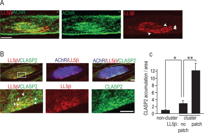
FIGURE 1:
LL5β colocalizes with CLASP2 at agrin-induced AChR clusters. (A) LL5β is enriched at agrin-induced AChR clusters. Primary myotubes were cultured on focal agrin deposits on a laminin substrate, and AChR clusters were stained with α-BTX–Alexa 488 (green) and for LL5β (red). Arrowheads mark uneven distribution of LL5β. Scale bar, 5 μm. (B) Regions of elevated LL5β (red) inside AChR clusters (blue) are enriched for CLASP2 (green, marked with arrowheads), consistent with an LL5β-CLASP2 interaction and CLASP2-dependent MT capture at synaptic membranes by LL5β. At 48 h postinfection, myotubes expressing adenovirus-delivered GFP-CLASP2 were stained for AChRs (blue), endogenous LL5β (red), and GFP with antibody. Bottom, magnification of boxed area at top. Scale bar, 10 μm (top), 5 μm (bottom). (C) Quantification of LL5β and CLASP2 inside AChR clusters. Bar graph shows that CLASP2 load/area increases with LL5β levels within the myotube region indicated on x-axis. Means ± SE; **p < 0.01; *p < 0.05; n = 6 myotubes with >10 comets/cell.

