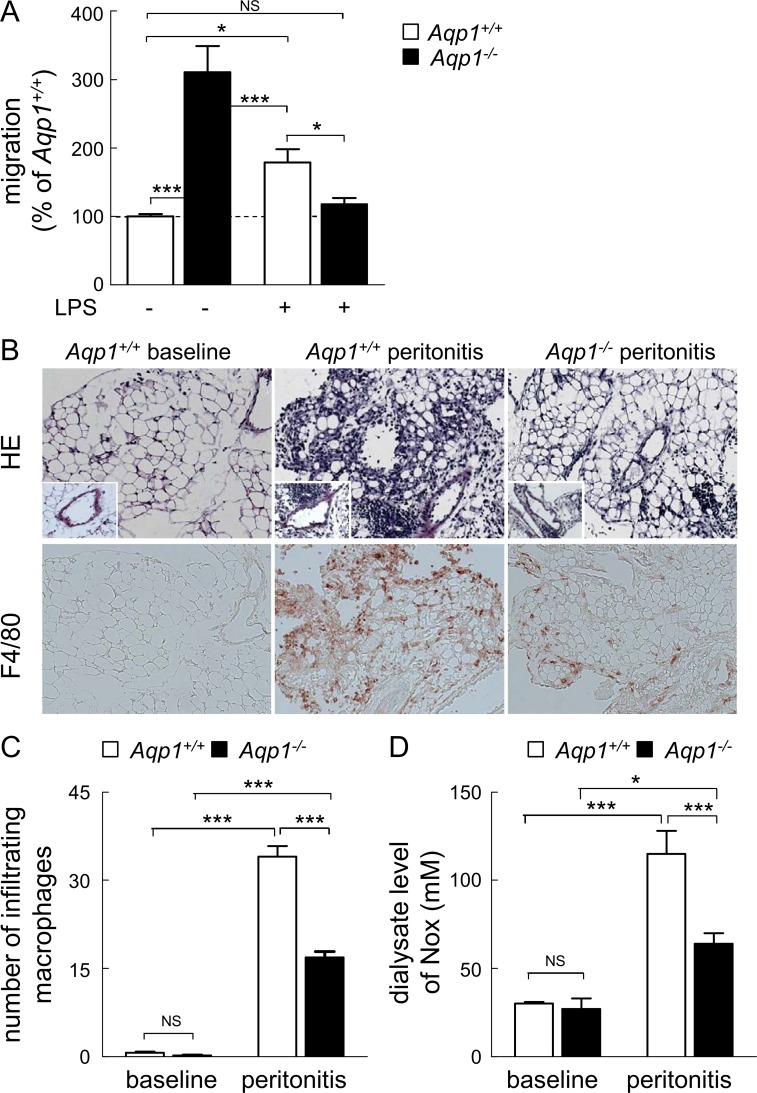
Fig 7. AQP1 ablation decreases macrophage migration upon LPS stimulation and in an inflammatory infiltrate.
(A) In vitro wound healing assay upon LPS stimulation. Migration of Aqp1 +/+ (open bars) or Aqp1 -/- (filled bars) macrophages was tested as in Fig. 1 in medium with serum alone (undifferentiated M0 phenotype) or supplemented by LPS (M1 phenotype). Values are expressed by reference to Aqp1 +/+ macrophages maintained in serum alone and are means±SEM of 4–6 experiments with 3 dishes each. (B-D) In vivo peritonitis model. The morphology (hematoxylin-eosin [HE] staining) and the presence of infiltrating macrophages (immunolabeling for F4/80) in the visceral peritoneum were assessed in Aqp1 +/+ and Aqp1 -/- mice at baseline and 7 days after generation of an acute peritonitis (B). The peritoneal infiltrate as well as the number of infiltrating macrophages are greatly attenuated in Aqp1 -/- vs Aqp1 +/+ mice, as confirmed by the quantification of infiltrating macrophages (C) and dialysate level of NOx (D). Original magnification, x150 (inset, x300). Representative of 4 trios of mice. NS, not significant; *, p<0.05; ***, p<0.001.

