
FIGURE 2.
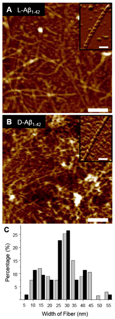
The (A) L- and (B) D-Aβ1-42 isomers induced towards fibril formation in the absence of a lipid membrane by incubation in 1% NH4OH at 37 °C for 72 h, dried overnight on fresh mica, and imaged with AFM in air. Both isomers form similar complex networks of globular units, oligomers, protofibrils, and fibrils. Insets show high resolution images of individual fibers (scale bars = 250 nm, height color scales = 25 nm, inset scale bars = 100 nm). (C) The distribution of randomly measured widths (n = 67, here n denotes the number of samples) of fibers of the D-Aβ1-42 fibers (gray bars) after incubation at 37 °C for 72 h shows a distribution with fiber widths most frequently between 25 nm and 30 nm. The distribution of the L-Aβ1-42 fibril widths (black bars), after similar incubation, also shows a distribution (n = 53) with fiber widths most frequently between 25 nm and 30nm. The overall range of values is comparable for both isomers.