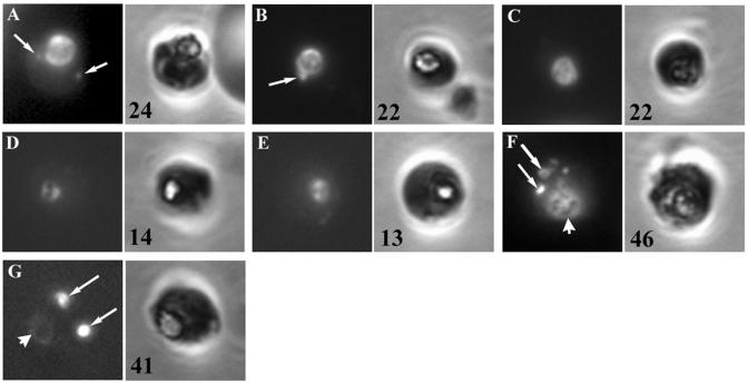
FIG. 6.
RBC-free endocytosis assay. Parasites released from cultured pRBCs by saponin treatment were incubated for 5 h in a diluted RBC lysate containing FITC-dextran and no drug (A), 120 nM chloroquine (B), 1.5 μM chloroquine (C), mefloquine (D), artemisinin (E), ammonium chloride (F), or monensin (G). The parasites were washed, fixed, and viewed by fluorescence microscopy. Left panels, fluorescence images of individual parasites; right panels, the corresponding phase-contrast images. Arrows, transport vesicles; arrowheads, positions of the food vacuole. The percentages of parasites containing two or more transport vesicles are indicated in the bottom left corner of the phase-contrast panels.

