
Figure 3.
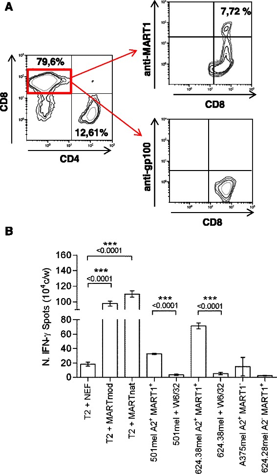
Phenotypic and functional analysis of tumor antigen-specific CD8 T cells. (A) Phenotypic analysis of pentamer+ CD8+ T cells after sensitization with the HLA2-A*0201 restricted-modified peptides (Melan-A/MART-1[27L] or gp100[210M]). (B) The tumor specificity of peptide sensitized T cells was assessed by measuring IFN-γ secretion (Enzyme-Linked ImmunoSpot (ELISpot) assay) upon stimulation with HLA-A*0201-restricted Melan A/MART-1 (modified or native)-pulsed (2 μmol/L) lymphoblastoid T2 cell line or HLA-matched HLA-A*0201+MART1+ tumor cells (#501mel and #624.38mel) pretreated or not with the anti-HLA class I (W6/32) mAb. Moreover, T cells were also incubated with HLA-mismatched allogeneic HLA-A*0201−MART1+ (#624.28mel) or HLA-A*0201+MART1− melanoma cells (#A375mel). The irrelevant peptide NEF[180–189] was used as negative control. P values were calculated by two-tailed t test.