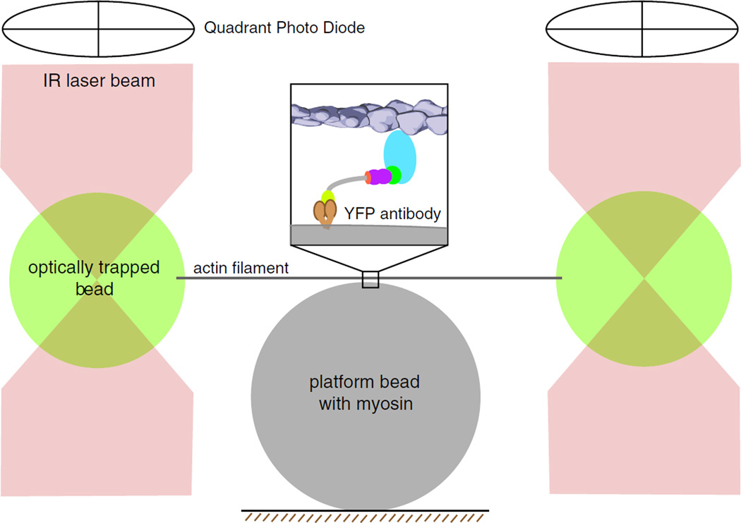
Fig. 4.
Optical trapping of myosin molecules: cartoon schematic of a dualbeam laser optical trap for measuring the lever-arm stroke, force generation, and kinetics of single myosin molecules. Streptavidin-coated polystyrene beads are optically trapped by a focused infrared (IR) laser beam. An optically trapped bead is attached to each end of a single biotinylated actin filament (gray line). The filament is stabilized using fluorescently labeled phalloidin. The actin filament is stretched taut in the optical trap by independently steering the two laser beams. Inset A myosin VI monomeric construct with C-terminal GFP tags is attached to polystyrene “platform” beads coated with anti-GFP antibody. The position of each of the trap beads is determined accurately by two quadrant photo detectors (QPD) positioned above the optical trap. QPD readout can be calibrated to yield an accurate measure of myosin step size

