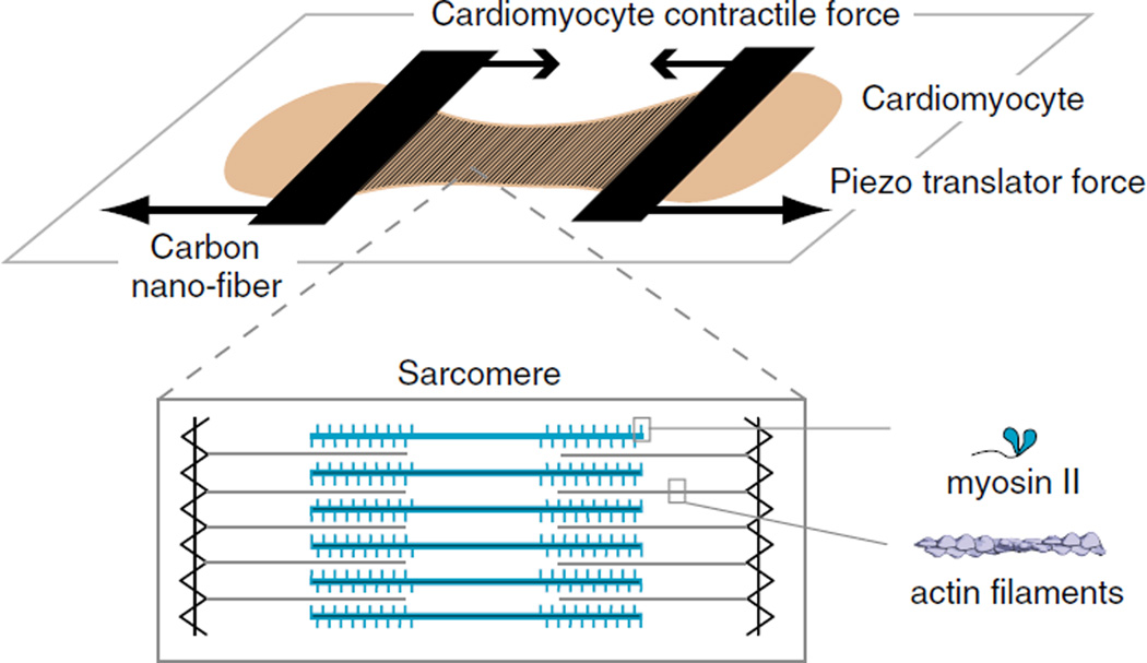
Fig. 5.
Single cardiomyocyte dynamic force–length measurements: cartoon schematic of a cardiomyocyte (light brown) suspended between two carbon nanofibers. The carbon nanofibers act as calibrated force transducers, whose displacement is a dynamic readout of the force generated by the cardiomyocyte. The carbon nanofibers can be coupled to a piezo-translator. This provides a high-frequency feedback that adjusts the position of the carbon nanofiber during cardiomyocyte contraction, thereby simulating physiological load conditions. Inset The contractile apparatus of a cardiomyocyte, namely, the muscle sarcomere, is shown, highlighting the spatial arrangement of actin filaments (gray) and myosin thick filaments (blue), which are integrated with numerous structural and regulatory proteins (not shown) to generate a contractile force

