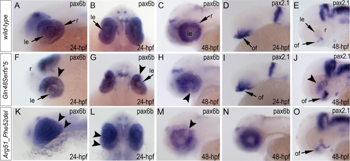
Fig 7. Analysis of pax6b, pax2.1 and foxe3 expression in wild-type, mab21l2 Q48Sfs*5 and mab21l2 R51_F52del embryos.
Wild-type (A-E) and mutant (F-O) zebrafish embryos at 24–48-hpf were analyzed as indicated in the right bottom corner of each image. Please note a change in pax6b transcript distribution at 24-hpf and 48-hpf in mab21l2 Q48Sfs*5 (arrowheads in F-H), retinal folding defect in mab21l2 R51_F52del embryos at 24-hpf (arrowheads in K, L) and visibly abnormal pax6b pattern at 48-hpf in some (arrowheads in M) but not all (N) mab21l2 R51_F52del embryos. pax2.1 expression seems to be unaffected in 24-hpf frameshift mutant embryos (I) but shows an abnormal pattern in both mutants at 48-hpf (J,O). At 48-hpf, in addition to more broad and intense pax2.1 expression in the region of optic fissure, abnormal pax2.1 staining was detected in central retina in mab21l2 Q48Sfs*5 embryos (arrowheads in J). le, lens; of, optic fissure; retina.

